This blog posts explains how to train a deep learning lymphoma sub-type classifier in accordance with our paper “Deep learning for digital pathology image analysis: A comprehensive tutorial with selected use cases”.
This blog posts explains how to train a deep learning mitosis detector in accordance with our paper “Deep learning for digital pathology image analysis: A comprehensive tutorial with selected use cases”.
This blog posts explains how to train a deep learning Invasive Ductal Carcinoma (IDC) classifier in accordance with our paper “Deep learning for digital pathology image analysis: A comprehensive tutorial with selected use cases”.
This blog posts explains how to train a deep learning tubule segmentation classifier in accordance with our paper “Deep learning for digital pathology image analysis: A comprehensive tutorial with selected use cases”.
Typically, you’ll want to use a validation set to determine an optimal threshold as it is often not .5 (which is equivalent to argmax). Subsequently, use this threshold on the the “_prob” image to generate a binary image.This blog posts explains how to train a deep learning lymphocyte detector in accordance with our paper “Deep learning for digital pathology image analysis: A comprehensive tutorial with selected use cases”.
This blog posts explains how to train a deep learning epithelium segmentation classifier in accordance with our paper “Deep learning for digital pathology image analysis: A comprehensive tutorial with selected use cases”.
This blog posts explains how to train a deep learning nuclear segmentation classifier in accordance with our paper “Deep learning for digital pathology image analysis: A comprehensive tutorial with selected use cases”.
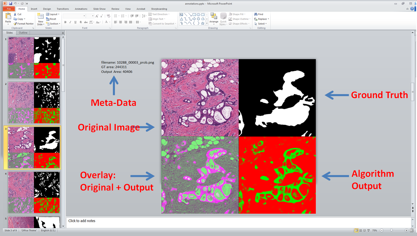
Reviewing the results of an image based experiment, across many images, can be annoying in matlab. Too much clicking!
I’ve recently started using PowerPoint to view many of my results. This blog posts discuss how using the free export to PowerPoint toolbox it is possible to create a slide desk with all relevant information for easier viewing. It looks like this:
One of the perks of working at Case Western Reserve is that we often qualify for access to cutting edge resource and special projects. In this case, since our digital histology deep learning work requires a large number of GPUs to analyze thousands of patients, we were granted access to the OSC Ruby cluster, which has 20 NVIDIA Tesla K40 GPUs. Since the cluster has only recently been setup, there was some leg work required on our end to get Caffe fully up and running, without root access, which we’ll document here.
Annotations which stay solely in Bisque aren’t incredibly useful. One of the main reasons for marking up images is to be able to use those annotations for some other purpose, such as training classifiers, computing metrics and features. In this post, we show how to take the annotations from bisque via REST, and convert them into binary masks in matlab.