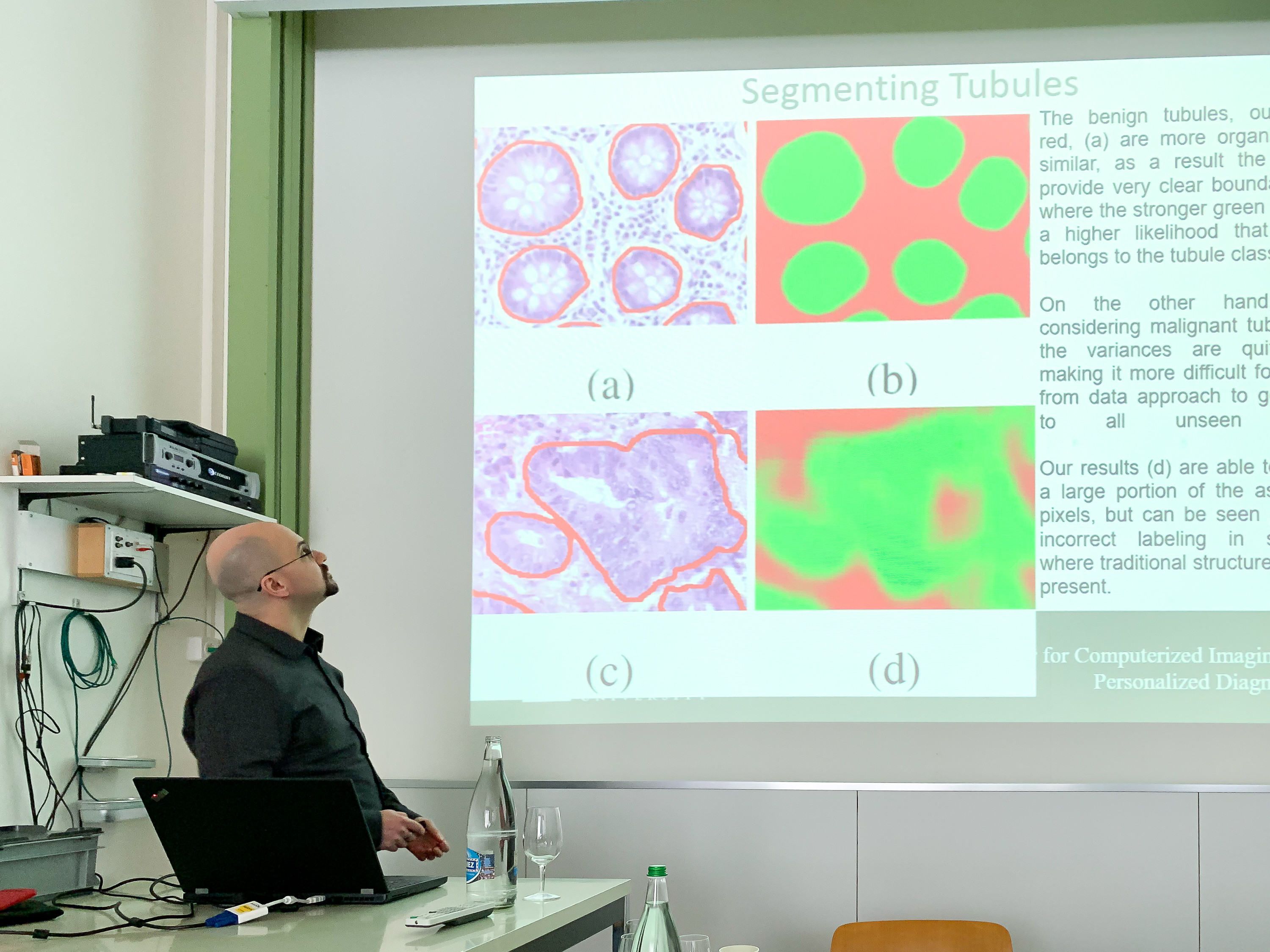
Thanks to everyone for their attendance in our full-day training course in Basel, Switzerland!


Deep learning (DL) models have been performing exceptionally well on a number of challenging tasks lately. Unfortunately, given the current blackbox nature of these DL models, it is difficult to try and “understand” what the network is seeing and how it is making its decisions. Building upon our previous post discussing how to train a DenseNet for classification, we discuss here how to apply various visualization techniques to enable us to interrogate the network. The code here is designed as drop-in functionality for any network trained using the previous post, hopefully easing the burden of its implementation.
In this blog post, we discuss how to train a DenseNet style deep learning classifier, using Pytorch, for differentiating between different types of lymphoma cancer. This post and code are based on the post discussing segmentation using U-Net and is thus broken down into the same 4 components:
Continue reading Digital pathology classification using Pytorch + Densenet
In this blog post, we discuss how to train a U-net style deep learning classifier, using Pytorch, for segmenting epithelium versus stroma regions. This post is broken down into 4 components following along other pipeline approaches we’ve discussed in the past:
This model focuses on using solely Python and freely available tools (i.e., no matlab).
This blog post assumes moderate knowledge of convolutional neural networks, depending on the readers background, our JPI paper may be sufficient, or a more thorough resource such as Andrew NG’s deep learning course.
Continue reading Digital Pathology Segmentation using Pytorch + Unet
As we’re testing out for migration to new deep learning frameworks, one of the questions that remained was dataset interoperability. Essentially, we want to be able to create a dataset for training a deep learning framework from as many applications as possible (python, matlab, R, etc), so that our students can use a language that are familiar to them, as well as leverage all of the existing in-house code we have for data manipulation.
Continue reading Using Matlab, Pytables (hdf5) and (a bit of) Pytorch
This is a very straightforward practical approach:
https://github.com/NVIDIA/DIGITS/tree/master/examples/fine-tuning
One trick I’ve learned from somewhere (can’t find the link, unfortunately), which is a break from the above tutorial, is to simply reduce the base learning rate by an order of magnitude when transferring, while simultaneously seting the “new” layers to have their lr_mult an order of magnitude higher than the rest of the network
so, to initially train:
to transfer learn:
This saves a lot of editing of the files and allows for small amounts of adjustment to existing layers, while focusing the bulk of the learning on the newer layers, yet still resisting to overfitting small datasets.
This is pretty informative:
https://cs231n.github.io/transfer-learning/
And I think this is the original TL paper as far as I remember:
Just wanted to take a moment and share some quick stain normalization type experimental results. We have a trained in-house nuclei segmentation model which works fairly well when the test images have similar stain presentation properties, but when new datasets arrive which are notably different we tend to see a decreased classifier performance.
Here we look at one of these images and ways of improving classifier robustness.
One of the common ways of increasing the size of a training set is to augment the original data with a set of modified patches. These modifications often include (a) rotations, (b) mirroring, (c) lighting adjustment, (d) affine transformations (sheering, etc), (e) magnification modification, (f) addition of noise, etc. This blog post discusses how to do the most trivial modification, rotation, in real-time using a python layer through Nvidia Digits. Given this code, it should be easy to add on other desired augmentations.
Continue reading Real time Data Augmentation using Nvidia Digits + Python Layer
Our publication “Deep learning for digital pathology image analysis: A comprehensive tutorial with selected use cases” , showed how to use deep learning to address many common digital pathology tasks. Since then, many improvements have been made both in the field and in my implementation of them. In this blog post, I re-address the nuclei segmentation use case using the latest and greatest approaches.
Continue reading Revised Deep Learning approach using Matlab + Caffe + Python
This tutorial provides a tutorial on using the code and data for our paper “A resolution adaptive deep hierarchical (RADHicaL) learning scheme applied to nuclear segmentation of digital pathology images” by Andrew Janowczyk, Scott Doyle, Hannah Gilmore, and Anant Madabhushi.

