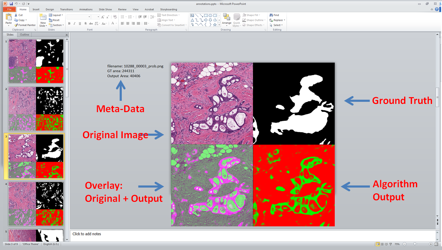
Reviewing the results of an image based experiment, across many images, can be annoying in matlab. Too much clicking!
I’ve recently started using PowerPoint to view many of my results. This blog posts discuss how using the free export to PowerPoint toolbox it is possible to create a slide desk with all relevant information for easier viewing. It looks like this:
Annotations which stay solely in Bisque aren’t incredibly useful. One of the main reasons for marking up images is to be able to use those annotations for some other purpose, such as training classifiers, computing metrics and features. In this post, we show how to take the annotations from bisque via REST, and convert them into binary masks in matlab.
Once we have images uploaded to Bisque, we might want to be able to overlay annotations on top of them so that users can interact with them. This post quickly goes through the conversation of a binary mask, in matlab, into an XML which can later be imported into Bisque.
In the previous post we discussed how to export annotations from a Ventana Image Viewer program and create binary masks. Now we explain how to do the opposite and import the mask back into Image Viewer.
There are often times when we want to see the boundaries of an annotation overlaid on an image for easier inspection. Using the ‘AlphaData’ layer in matlab this becomes extremely easy and efficient. Continue reading Overlaying Binary Masks on Images in Matlab→
One of the main purposes of having a digital format is to allow experts (e.g., pathologists) to annotate certain structures in the images. Be it nuclei, epithelium/stroma regions, tumor/non-tumor tissue etc. This is easily done with ImageScope and SVS files, but the trick is importing them into Matlab. Continue reading Working with Aperio SVS files in Matlab – Converting Annotations to Binary Masks→
In the previous tutorial we discussed how to load different levels of the image pyramid for Aperio SVS images in Matlab and how they corresponded.
In this tutorial we will extend upon that in 2 critical ways. First, we’d like to be able to load only small sub-sections from images (easy) and then extend upon that so we can identify regions at a low-magnification that we’d like to load the corresponding high-magnification version (harder). Continue reading Working with Aperio SVS files in Matlab – Loading Sub-Sections→
Aperio scanners generate a semi-proprietary file format called SVS. At its heart, SVS files are really a multi-page tiff file storing a pyramid of smaller tiff files of the original image. We’ll look at those here using a SVS file provided by the TCGA (http://cancergenome.nih.gov/) breast cancer cohort: